Cadherin-2
Protein-coding gene in the species Homo sapiens
| CDH2 | |||||||||||||||||||||||||||||||||||||||||||||||||||
|---|---|---|---|---|---|---|---|---|---|---|---|---|---|---|---|---|---|---|---|---|---|---|---|---|---|---|---|---|---|---|---|---|---|---|---|---|---|---|---|---|---|---|---|---|---|---|---|---|---|---|---|
 | |||||||||||||||||||||||||||||||||||||||||||||||||||
| Identifiers | |||||||||||||||||||||||||||||||||||||||||||||||||||
| Aliases | CDH2, CD325, CDHN, CDw325, NCAD, cadherin 2, ACOGS, ARVD14 | ||||||||||||||||||||||||||||||||||||||||||||||||||
| External IDs | OMIM: 114020; MGI: 88355; HomoloGene: 20424; GeneCards: CDH2; OMA:CDH2 - orthologs | ||||||||||||||||||||||||||||||||||||||||||||||||||
| |||||||||||||||||||||||||||||||||||||||||||||||||||
| |||||||||||||||||||||||||||||||||||||||||||||||||||
| |||||||||||||||||||||||||||||||||||||||||||||||||||
| |||||||||||||||||||||||||||||||||||||||||||||||||||
| |||||||||||||||||||||||||||||||||||||||||||||||||||
| Wikidata | |||||||||||||||||||||||||||||||||||||||||||||||||||
| |||||||||||||||||||||||||||||||||||||||||||||||||||


Cadherin-2 also known as Neural cadherin (N-cadherin), is a protein that in humans is encoded by the CDH2 gene.[5][6][7] CDH2 has also been designated as CD325 (cluster of differentiation 325). Cadherin-2 is a transmembrane protein expressed in multiple tissues and functions to mediate cell–cell adhesion. In cardiac muscle, Cadherin-2 is an integral component in adherens junctions residing at intercalated discs, which function to mechanically and electrically couple adjacent cardiomyocytes. Alterations in expression and integrity of Cadherin-2 has been observed in various forms of disease, including human dilated cardiomyopathy. Variants in CDH2 have also been identified to cause a syndromic neurodevelopmental disorder.[8]
Structure
Cadherin-2 is a protein with molecular weight of 99.7 kDa, and 906 amino acids in length.[9] Cadherin-2, a classical cadherin from the cadherin superfamily, is composed of five extracellular cadherin repeats, a transmembrane region and a highly conserved cytoplasmic tail. Cadherin-2, as well as other cadherins, interact with Cadherin-2 on an adjacent cell in an anti-parallel conformation, thus creating a linear, adhesive "zipper" between cells.[10]
Function
Cadherin-2, originally named Neural cadherin for its role in neural tissue, plays a role in neurons and later was found to also play a role in cardiac muscle and in cancer metastasis. Cadherin-2 is a transmembrane, homophilic glycoprotein belonging to the calcium-dependent cell adhesion molecule family. These proteins have extracellular domains that mediate homophilic interactions between adjacent cells, and C-terminal, cytoplasmic tails that mediate binding to catenins, which in turn interact with the actin cytoskeleton.[11][12][13]
Role in development
Cadherin-2 plays a role in development as a calcium dependent cell–cell adhesion glycoprotein that functions during gastrulation and is required for establishment of left-right asymmetry.[14]
Cadherin-2 is widely expressed in the embryo post-implantation, showing high levels in the mesoderm with sustained expression through adulthood.[15] Cadherin-2 mutation during development has the most significant effect on cell adhesion in the primitive heart; dissociated myocytes and abnormal heart tube development occur.[16] Cadherin-2 plays a role in the development of the vertebrate heart at the transition of epithelial cells to trabecular and compact myocardial cell layer formation.[17] An additional study showed that myocytes expressing a dominant negative Cadherin-2 mutant showed significant abnormalities in myocyte distribution and migration towards the endocardium, resulting in defects in trabecular formation within the myocardium.[18][19]
Role in cardiac muscle
In cardiac muscle, Cadherin-2 is found at intercalated disc structures which provide end-on cell–cell connections that facilitate mechanical and electrical coupling between adjacent cardiomyocytes. Within intercalated discs are three types of junctions: adherens junctions, desmosomes and gap junctions;[20] Cadherin-2 is an essential component in adherens junctions, which enables cell–cell adhesion and force transmission across the sarcolemma.[21] Cadherin-2 complexed to catenins has been described as a master regulator of intercalated disc function.[22] Cadherin-2 appears at cell–cell junctions prior to gap junction formation,[23][24] and is critical for normal myofibrillogenesis.[25] Expression of a mutant form of Cadherin-2 harboring a large deletion in the extracellular domain inhibited the function of endogenous Cadherin-2 in adult ventricular cardiomyocytes, and neighboring cardiomyocytes lost cell–cell contact and gap junction plaques as well.[26]
Mouse models employing transgenesis have highlighted the function of N-cadherin in cardiac muscle. Mice with altered expression of N-cadherin and/or E-cadherin showed a dilated cardiomyopathy phenotype, likely due to malfunction of intercalated discs.[27] In agreement with this, mice with ablation of N-cadherin in adult hearts via a cardiac-specific tamoxifen-inducible Cre N-cadherin transgene showed disrupted assembly of intercalated discs, dilated cardiomyopathy, impaired cardiac function, decreased sarcomere length, increased Z-line thickness, decreases in connexin 43, and a loss in muscular tension. Mice died within two months of transgene expression, mainly due to spontaneous Ventricular tachycardia.[28] Further analysis of N-cadherin knockout mice revealed that the arrhythmias were likely due to ion channel remodeling and aberrant Kv1.5 channel function. These animals showed a prolonged action potential duration, reduced density of inward rectifier potassium channel and decreased expression of Kv1.5, KCNE2 and cortactin combined with disrupted actin cytoskeleton at the sarcolemma.[29]
Role in neurons
In neural cells, at certain central nervous system synapses, presynaptic to postsynaptic adhesion is mediated at least in part by Cadherin-2.[30] N-cadherins interact with catenins to play an important role in learning and memory (For full article see Cadherin-catenin complex in learning and memory). Loss of N-cadherin is also associated with attention-deficit hyperactivity disorder in humans, and impaired synaptic functioning. [31]
Role in cancer metastasis
Cadherin-2 is commonly found in cancer cells and provides a mechanism for transendothelial migration. When a cancer cell adheres to the endothelial cells of a blood vessel it up-regulates the src kinase pathway, which phosphorylates beta-catenins attached to both Cadherin-2 (this protein) and E-cadherins. This causes the intercellular connection between two adjacent endothelial cells to fail and allows the cancer cell to slip through.[32]
Clinical significance
Variants in CDH2 have been identified to cause a syndromic neurodevelopmental disorder characterized by Corpus callosum, axon, cardiac, ocular, and genital differences.[8]
One study investigating genetic underpinnings of obsessive-compulsive disorder and Tourette disorder found that while CDH2 variants are likely not disease-causing as single entities, they may confer risk when examined as part of a panel of related cell–cell adhesion genes.[33] Further studies in larger cohorts will be required to unequivocally determine this.
In human dilated cardiomyopathy, it was shown that Cadherin-2 expression was enhanced and arranged in a disarrayed fashion, suggesting that disorganization of Cadherin-2 protein in heart disease may be a component of remodeling.[34]
Interactions
Cadherin-2 has been shown to interact with:
See also
References
- ^ a b c GRCh38: Ensembl release 89: ENSG00000170558 – Ensembl, May 2017
- ^ a b c GRCm38: Ensembl release 89: ENSMUSG00000024304 – Ensembl, May 2017
- ^ "Human PubMed Reference:". National Center for Biotechnology Information, U.S. National Library of Medicine.
- ^ "Mouse PubMed Reference:". National Center for Biotechnology Information, U.S. National Library of Medicine.
- ^ "UniProt". www.uniprot.org. Retrieved 26 August 2022.
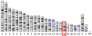
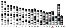
- ^ Walsh FS, Barton CH, Putt W, Moore SE, Kelsell D, Spurr N, Goodfellow PN (September 1990). "N-cadherin gene maps to human chromosome 18 and is not linked to the E-cadherin gene". Journal of Neurochemistry. 55 (3): 805–12. doi:10.1111/j.1471-4159.1990.tb04563.x. PMID 2384753. S2CID 29840435.
- ^ Reid RA, Hemperly JJ (October 1990). "Human N-cadherin: nucleotide and deduced amino acid sequence". Nucleic Acids Research. 18 (19): 5896. doi:10.1093/nar/18.19.5896. PMC 332345. PMID 2216790.
- ^ a b Accogli A, Calabretta S, St-Onge J, Boudrahem-Addour N, Dionne-Laporte A, Joset P, et al. (October 2019). "De Novo Pathogenic Variants in N-cadherin Cause a Syndromic Neurodevelopmental Disorder with Corpus Collosum, Axon, Cardiac, Ocular, and Genital Defects". American Journal of Human Genetics. 105 (4): 854–868. doi:10.1016/j.ajhg.2019.09.005. PMC 6817525. PMID 31585109.
- ^ "Protein sequence of human CDH2 (Uniprot ID: P19022)". Cardiac Organellar Protein Atlas Knowledgebase (COPaKB). Archived from the original on 24 September 2015. Retrieved 20 July 2015.
- ^ Shapiro L, Fannon AM, Kwong PD, Thompson A, Lehmann MS, Grübel G, et al. (March 1995). "Structural basis of cell-cell adhesion by cadherins". Nature. 374 (6520): 327–37. Bibcode:1995Natur.374..327S. doi:10.1038/374327a0. PMID 7885471. S2CID 4314442.
- ^ Buxton RS, Magee AI (June 1992). "Structure and interactions of desmosomal and other cadherins". Seminars in Cell Biology. 3 (3): 157–67. doi:10.1016/s1043-4682(10)80012-1. PMID 1623205.
- ^ Takeichi M (1990). "Cadherins: a molecular family important in selective cell-cell adhesion". Annual Review of Biochemistry. 59: 237–52. doi:10.1146/annurev.bi.59.070190.001321. PMID 2197976.
- ^ Ozawa M, Baribault H, Kemler R (June 1989). "The cytoplasmic domain of the cell adhesion molecule uvomorulin associates with three independent proteins structurally related in different species". The EMBO Journal. 8 (6): 1711–7. doi:10.1002/j.1460-2075.1989.tb03563.x. PMC 401013. PMID 2788574.
- ^ García-Castro MI, Vielmetter E, Bronner-Fraser M (May 2000). "N-Cadherin, a cell adhesion molecule involved in establishment of embryonic left-right asymmetry". Science. 288 (5468): 1047–51. Bibcode:2000Sci...288.1047G. doi:10.1126/science.288.5468.1047. PMID 10807574.
- ^ Angst BD, Khan LU, Severs NJ, Whitely K, Rothery S, Thompson RP, et al. (January 1997). "Dissociated spatial patterning of gap junctions and cell adhesion junctions during postnatal differentiation of ventricular myocardium". Circulation Research. 80 (1): 88–94. doi:10.1161/01.res.80.1.88. PMID 8978327.
- ^ Radice GL, Rayburn H, Matsunami H, Knudsen KA, Takeichi M, Hynes RO (January 1997). "Developmental defects in mouse embryos lacking N-cadherin". Developmental Biology. 181 (1): 64–78. doi:10.1006/dbio.1996.8443. PMID 9015265.
- ^ Kostetskii I, Moore R, Kemler R, Radice GL (June 2001). "Differential adhesion leads to segregation and exclusion of N-cadherin-deficient cells in chimeric embryos". Developmental Biology. 234 (1): 72–9. doi:10.1006/dbio.2001.0250. PMID 11356020.
- ^ Linask KK, Knudsen KA, Gui YH (May 1997). "N-cadherin-catenin interaction: necessary component of cardiac cell compartmentalization during early vertebrate heart development". Developmental Biology. 185 (2): 148–64. doi:10.1006/dbio.1997.8570. PMID 9187080.
- ^ Ong LL, Kim N, Mima T, Cohen-Gould L, Mikawa T (January 1998). "Trabecular myocytes of the embryonic heart require N-cadherin for migratory unit identity". Developmental Biology. 193 (1): 1–9. doi:10.1006/dbio.1997.8775. PMID 9466883.
- ^ Peters NS, Severs NJ, Rothery SM, Lincoln C, Yacoub MH, Green CR (August 1994). "Spatiotemporal relation between gap junctions and fascia adherens junctions during postnatal development of human ventricular myocardium". Circulation. 90 (2): 713–25. doi:10.1161/01.cir.90.2.713. PMID 8044940.
- ^ Forbes MS, Sperelakis N (1985). "Intercalated discs of mammalian heart: a review of structure and function". Tissue & Cell. 17 (5): 605–48. doi:10.1016/0040-8166(85)90001-1. PMID 3904080.
- ^ Vite A, Radice GL (June 2014). "N-cadherin/catenin complex as a master regulator of intercalated disc function". Cell Communication & Adhesion. 21 (3): 169–79. doi:10.3109/15419061.2014.908853. PMC 6054126. PMID 24766605.
- ^ Zuppinger C, Schaub MC, Eppenberger HM (April 2000). "Dynamics of early contact formation in cultured adult rat cardiomyocytes studied by N-cadherin fused to green fluorescent protein". Journal of Molecular and Cellular Cardiology. 32 (4): 539–55. doi:10.1006/jmcc.1999.1086. PMID 10756112.
- ^ Dou JP, Jiao B, Sheng JJ, Yu ZB (October 2014). "[Dynamic assembly of intercalated disc during postnatal development in the rat myocardium]". Sheng Li Xue Bao. 66 (5): 569–74. PMID 25332002.
- ^ Goncharova EJ, Kam Z, Geiger B (January 1992). "The involvement of adherens junction components in myofibrillogenesis in cultured cardiac myocytes". Development. 114 (1): 173–83. doi:10.1242/dev.114.1.173. PMID 1576958.
- ^ Hertig CM, Eppenberger-Eberhardt M, Koch S, Eppenberger HM (January 1996). "N-cadherin in adult rat cardiomyocytes in culture. I. Functional role of N-cadherin and impairment of cell-cell contact by a truncated N-cadherin mutant". Journal of Cell Science. 109 ( Pt 1) (1): 1–10. doi:10.1242/jcs.109.1.1. PMID 8834785.
- ^ Ferreira-Cornwell MC, Luo Y, Narula N, Lenox JM, Lieberman M, Radice GL (April 2002). "Remodeling the intercalated disc leads to cardiomyopathy in mice misexpressing cadherins in the heart". Journal of Cell Science. 115 (Pt 8): 1623–34. doi:10.1242/jcs.115.8.1623. PMID 11950881.
- ^ Kostetskii I, Li J, Xiong Y, Zhou R, Ferrari VA, Patel VV, et al. (February 2005). "Induced deletion of the N-cadherin gene in the heart leads to dissolution of the intercalated disc structure". Circulation Research. 96 (3): 346–54. doi:10.1161/01.RES.0000156274.72390.2c. PMID 15662031.
- ^ Cheng L, Yung A, Covarrubias M, Radice GL (June 2011). "Cortactin is required for N-cadherin regulation of Kv1.5 channel function". The Journal of Biological Chemistry. 286 (23): 20478–89. doi:10.1074/jbc.m111.218560. PMC 3121477. PMID 21507952.
- ^ "Entrez Gene: CDH2 cadherin 2, type 1, N-cadherin (neuronal)".
- ^ Halperin D, Stavsky A, Kadir R, Drabkin M, Wormser O, Yogev Y (October 2021). "CDH2 mutation affecting N-cadherin function causes attention-deficit hyperactivity disorder in humans and mice". Nature Communications. 6187 (12): 625–30. Bibcode:2021NatCo..12.6187H. doi:10.1038/s41467-021-26426-1. PMC 8548587. PMID 34702855.
- ^ Ramis-Conde I, Chaplain MA, Anderson AR, Drasdo D (March 2009). "Multi-scale modelling of cancer cell intravasation: the role of cadherins in metastasis". Physical Biology. 6 (1): 016008. Bibcode:2009PhBio...6a6008R. doi:10.1088/1478-3975/6/1/016008. PMID 19321920. S2CID 206096620.
- ^ Moya PR, Dodman NH, Timpano KR, Rubenstein LM, Rana Z, Fried RL, et al. (August 2013). "Rare missense neuronal cadherin gene (CDH2) variants in specific obsessive-compulsive disorder and Tourette disorder phenotypes". European Journal of Human Genetics. 21 (8): 850–4. doi:10.1038/ejhg.2012.245. PMC 3722668. PMID 23321619.
- ^ Tsipis A, Athanassiadou AM, Athanassiadou P, Kavantzas N, Agrogiannis G, Patsouris E (September 2010). "Apoptosis-related factors p53, bcl-2 and the defects of force transmission in dilated cardiomyopathy". Pathology, Research and Practice. 206 (9): 625–30. doi:10.1016/j.prp.2010.05.007. PMID 20591580.
- ^ a b c d e Straub BK, Boda J, Kuhn C, Schnoelzer M, Korf U, Kempf T, et al. (December 2003). "A novel cell-cell junction system: the cortex adhaerens mosaic of lens fiber cells". Journal of Cell Science. 116 (Pt 24): 4985–95. doi:10.1242/jcs.00815. PMID 14625392.
- ^ a b c Wahl JK, Kim YJ, Cullen JM, Johnson KR, Wheelock MJ (May 2003). "N-cadherin-catenin complexes form prior to cleavage of the proregion and transport to the plasma membrane". The Journal of Biological Chemistry. 278 (19): 17269–76. doi:10.1074/jbc.M211452200. PMID 12604612.
- ^ Izawa I, Nishizawa M, Ohtakara K, Inagaki M (February 2002). "Densin-180 interacts with delta-catenin/neural plakophilin-related armadillo repeat protein at synapses". The Journal of Biological Chemistry. 277 (7): 5345–50. doi:10.1074/jbc.M110052200. PMID 11729199.
- ^ Brady-Kalnay SM, Rimm DL, Tonks NK (August 1995). "Receptor protein tyrosine phosphatase PTPmu associates with cadherins and catenins in vivo". The Journal of Cell Biology. 130 (4): 977–86. doi:10.1083/jcb.130.4.977. PMC 2199947. PMID 7642713.
- ^ Brady-Kalnay SM, Mourton T, Nixon JP, Pietz GE, Kinch M, Chen H, et al. (April 1998). "Dynamic interaction of PTPmu with multiple cadherins in vivo". The Journal of Cell Biology. 141 (1): 287–96. doi:10.1083/jcb.141.1.287. PMC 2132733. PMID 9531566.
- ^ Besco JA, Hooft van Huijsduijnen R, Frostholm A, Rotter A (October 2006). "Intracellular substrates of brain-enriched receptor protein tyrosine phosphatase rho (RPTPrho/PTPRT)". Brain Research. 1116 (1): 50–7. doi:10.1016/j.brainres.2006.07.122. PMID 16973135. S2CID 23343123.
- ^ Sacco PA, McGranahan TM, Wheelock MJ, Johnson KR (August 1995). "Identification of plakoglobin domains required for association with N-cadherin and alpha-catenin". The Journal of Biological Chemistry. 270 (34): 20201–6. doi:10.1074/jbc.270.34.20201. PMID 7650039.
- ^ Sinn HW, Balsamo J, Lilien J, Lin JJ (September 2002). "Localization of the novel Xin protein to the adherens junction complex in cardiac and skeletal muscle during development". Developmental Dynamics. 225 (1): 1–13. doi:10.1002/dvdy.10131. PMID 12203715. S2CID 23393425.
- ^ Schroen B, Leenders JJ, van Erk A, Bertrand AT, van Loon M, van Leeuwen RE, et al. (May 2007). "Lysosomal integral membrane protein 2 is a novel component of the cardiac intercalated disc and vital for load-induced cardiac myocyte hypertrophy". The Journal of Experimental Medicine. 204 (5): 1227–35. doi:10.1084/jem.20070145. PMC 2118572. PMID 17485520.
Further reading
- Doherty P, Smith P, Walsh FS (1997). "Shared cell adhesion molecule (CAM) homology domains point to CAMs signalling via FGF receptors". Perspectives on Developmental Neurobiology. 4 (2–3): 157–68. PMID 9168198.
- Makrigiannakis A, Coukos G, Blaschuk O, Coutifaris C (2000). "Follicular atresia and luteolysis. Evidence of a role for N-cadherin". Annals of the New York Academy of Sciences. 900 (1): 46–55. Bibcode:2000NYASA.900...46M. doi:10.1111/j.1749-6632.2000.tb06215.x. PMID 10818391. S2CID 30583247.
- Hazan RB, Qiao R, Keren R, Badano I, Suyama K (April 2004). "Cadherin switch in tumor progression". Annals of the New York Academy of Sciences. 1014 (1): 155–63. Bibcode:2004NYASA1014..155H. doi:10.1196/annals.1294.016. PMID 15153430. S2CID 37486403.
- Cavallaro U (December 2004). "N-cadherin as an invasion promoter: a novel target for antitumor therapy?". Current Opinion in Investigational Drugs. 5 (12): 1274–8. PMID 15648948.
- Salomon D, Ayalon O, Patel-King R, Hynes RO, Geiger B (May 1992). "Extrajunctional distribution of N-cadherin in cultured human endothelial cells". Journal of Cell Science. 102 ( Pt 1) (1): 7–17. doi:10.1242/jcs.102.1.7. PMID 1500442.
- Knudsen KA, Wheelock MJ (August 1992). "Plakoglobin, or an 83-kD homologue distinct from beta-catenin, interacts with E-cadherin and N-cadherin". The Journal of Cell Biology. 118 (3): 671–9. doi:10.1083/jcb.118.3.671. PMC 2289540. PMID 1639850.
- Reid RA, Hemperly JJ (October 1990). "Human N-cadherin: nucleotide and deduced amino acid sequence". Nucleic Acids Research. 18 (19): 5896. doi:10.1093/nar/18.19.5896. PMC 332345. PMID 2216790.
- Walsh FS, Barton CH, Putt W, Moore SE, Kelsell D, Spurr N, Goodfellow PN (September 1990). "N-cadherin gene maps to human chromosome 18 and is not linked to the E-cadherin gene". Journal of Neurochemistry. 55 (3): 805–12. doi:10.1111/j.1471-4159.1990.tb04563.x. PMID 2384753. S2CID 29840435.
- Selig S, Bruno S, Scharf JM, Wang CH, Vitale E, Gilliam TC, Kunkel LM (April 1995). "Expressed cadherin pseudogenes are localized to the critical region of the spinal muscular atrophy gene". Proceedings of the National Academy of Sciences of the United States of America. 92 (9): 3702–6. Bibcode:1995PNAS...92.3702S. doi:10.1073/pnas.92.9.3702. PMC 42029. PMID 7731968.
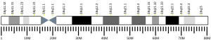
- Wallis J, Fox MF, Walsh FS (July 1994). "Structure of the human N-cadherin gene: YAC analysis and fine chromosomal mapping to 18q11.2". Genomics. 22 (1): 172–9. doi:10.1006/geno.1994.1358. PMID 7959764.
- Andersson AM, Edvardsen K, Skakkebaek NE (August 1994). "Expression and localization of N- and E-cadherin in the human testis and epididymis". International Journal of Andrology. 17 (4): 174–80. doi:10.1111/j.1365-2605.1994.tb01239.x. PMID 7995652.
- Matsuyoshi N, Imamura S (June 1997). "Multiple cadherins are expressed in human fibroblasts". Biochemical and Biophysical Research Communications. 235 (2): 355–8. doi:10.1006/bbrc.1997.6707. PMID 9199196.
- Navarro P, Ruco L, Dejana E (March 1998). "Differential localization of VE- and N-cadherins in human endothelial cells: VE-cadherin competes with N-cadherin for junctional localization". The Journal of Cell Biology. 140 (6): 1475–84. doi:10.1083/jcb.140.6.1475. PMC 2132661. PMID 9508779.
- Gaidar YA, Lepekhin EA, Sheichetova GA, Witt M (February 1998). "Distribution of N-cadherin and NCAM in neurons and endocrine cells of the human embryonic and fetal gastroenteropancreatic system". Acta Histochemica. 100 (1): 83–97. doi:10.1016/s0065-1281(98)80008-1. PMID 9542583.
- Kremmidiotis G, Baker E, Crawford J, Eyre HJ, Nahmias J, Callen DF (May 1998). "Localization of human cadherin genes to chromosome regions exhibiting cancer-related loss of heterozygosity". Genomics. 49 (3): 467–71. doi:10.1006/geno.1998.5281. PMID 9615235.
- Lu Q, Paredes M, Medina M, Zhou J, Cavallo R, Peifer M, et al. (February 1999). "delta-catenin, an adhesive junction-associated protein which promotes cell scattering". The Journal of Cell Biology. 144 (3): 519–32. doi:10.1083/jcb.144.3.519. PMC 2132907. PMID 9971746.
- Shan WS, Tanaka H, Phillips GR, Arndt K, Yoshida M, Colman DR, Shapiro L (February 2000). "Functional cis-heterodimers of N- and R-cadherins". The Journal of Cell Biology. 148 (3): 579–90. doi:10.1083/jcb.148.3.579. PMC 2174798. PMID 10662782.
- Husi H, Ward MA, Choudhary JS, Blackstock WP, Grant SG (July 2000). "Proteomic analysis of NMDA receptor-adhesion protein signaling complexes". Nature Neuroscience. 3 (7): 661–9. doi:10.1038/76615. hdl:1842/742. PMID 10862698. S2CID 14392630.
External links
- CDH2+protein,+human at the U.S. National Library of Medicine Medical Subject Headings (MeSH)
- CDH2 human gene location in the UCSC Genome Browser.
- CDH2 human gene details in the UCSC Genome Browser.
This article incorporates text from the United States National Library of Medicine, which is in the public domain.
- v
- t
- e
PDB gallery
-
 1ncg: STRUCTURAL BASIS OF CELL-CELL ADHESION BY CADHERINS
 -
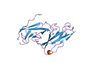 1nch: STRUCTURAL BASIS OF CELL-CELL ADHESION BY CADHERINS
 -
 1nci: STRUCTURAL BASIS OF CELL-CELL ADHESION BY CADHERINS
 -
 1ncj: N-CADHERIN, TWO-DOMAIN FRAGMENT


