Endolymphatic duct
Canal
| Endolymphatic duct | |
|---|---|
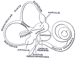 The membranous labyrinth. (Ductus endolymphaticus labeled at bottom center.) | |
 Endolymphatic duct is #6, and is labeled at top center. | |
| Details | |
| Identifiers | |
| Latin | ductus endolymphaticus |
| MeSH | D004711 |
| TA98 | A15.3.03.079 |
| TA2 | 7006 |
| FMA | 61246 |
| Anatomical terminology [edit on Wikidata] | |
From the posterior wall of the saccule a canal, the endolymphatic duct, is given off; this duct is joined by the ductus utriculosaccularis, and then passes along the aquaeductus vestibuli and ends in a blind pouch (endolymphatic sac) on the posterior surface of the petrous portion of the temporal bone, where it is in contact with the dura mater.
Disorders of the endolymphatic duct include Meniere's Disease and Enlarged Vestibular Aqueduct.
Additional images
-
 Transverse section through head of fetal sheep, in the region of the labyrinth. X 30.
 -
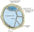 Transverse section of a human semicircular canal and duct


References
![]() This article incorporates text in the public domain from page 1052 of the 20th edition of Gray's Anatomy (1918)
This article incorporates text in the public domain from page 1052 of the 20th edition of Gray's Anatomy (1918)
External links
- The Endolymphatic Duct and Sac
- v
- t
- e
Anatomy of hearing and balance
- Auricle
- helix
- antihelix
- tragus
- antitragus
- intertragic notch
- earlobe
- Ear canal
- Auricular muscles
- Eardrum
| Tympanic cavity |
|
|---|---|
| Ossicles | |
| Auditory tube / Eustachian tube |
| Labyrinths | |||||
|---|---|---|---|---|---|
| Auditory system |
| ||||
| Vestibular system |
|
Portal:
 Anatomy

| Authority control databases |
|
|---|
 | This anatomy article is a stub. You can help Wikipedia by expanding it. |
- v
- t
- e


