Protein
| CDKN1A |
|---|
 |
| Available structures |
|---|
| PDB | Ortholog search: PDBe RCSB |
|---|
| List of PDB id codes |
|---|
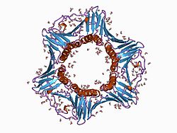
1AXC, 2ZVV, 2ZVW, 4RJF, 5E0U |
|
|
| Identifiers |
|---|
| Aliases | CDKN1A, CAP20, CDKN1, CIP1, MDA-6, P21, SDI1, WAF1, p21CIP1, cyclin-dependent kinase inhibitor 1A, cyclin dependent kinase inhibitor 1A |
|---|
| External IDs | OMIM: 116899; MGI: 104556; HomoloGene: 333; GeneCards: CDKN1A; OMA:CDKN1A - orthologs |
|---|
| Gene location (Human) |
|---|
 | | Chr. | Chromosome 6 (human)[1] |
|---|
| | Band | 6p21.2 | Start | 36,676,460 bp[1] |
|---|
| End | 36,687,339 bp[1] |
|---|
|
| Gene location (Mouse) |
|---|
 | | Chr. | Chromosome 17 (mouse)[2] |
|---|
| | Band | 17 A3.3|17 15.12 cM | Start | 29,309,950 bp[2] |
|---|
| End | 29,319,701 bp[2] |
|---|
|
| RNA expression pattern |
|---|
| Bgee | | Human | Mouse (ortholog) |
|---|
| Top expressed in | - stromal cell of endometrium
- gastric mucosa
- vena cava
- left uterine tube
- beta cell
- ascending aorta
- saphenous vein
- popliteal artery
- tibial arteries
- Descending thoracic aorta
|
| | Top expressed in | - stroma of bone marrow
- calvaria
- molar
- pyloric antrum
- lip
- esophagus
- hair follicle
- epithelium of stomach
- skin of external ear
- endothelial cell of lymphatic vessel
|
| | More reference expression data |
|
|---|
| BioGPS |  | | More reference expression data |
|
|---|
|
| Gene ontology |
|---|
| Molecular function | - metal ion binding
- protein binding
- cyclin-dependent protein serine/threonine kinase inhibitor activity
- ubiquitin protein ligase binding
- cyclin binding
- cyclin-dependent protein kinase activating kinase activity
- cyclin-dependent protein serine/threonine kinase activity
- protein kinase inhibitor activity
- protein kinase binding
- protein-containing complex binding
| | Cellular component | - cytoplasm
- cytosol
- cyclin-dependent protein kinase holoenzyme complex
- PCNA-p21 complex
- perinuclear region of cytoplasm
- nucleus
- nucleoplasm
- nucleolus
- nuclear body
- protein-containing complex
| | Biological process | - cellular response to extracellular stimulus
- signal transduction by p53 class mediator
- intrinsic apoptotic signaling pathway in response to DNA damage by p53 class mediator
- cellular response to heat
- regulation of cyclin-dependent protein serine/threonine kinase activity
- DNA damage response, signal transduction by p53 class mediator resulting in cell cycle arrest
- positive regulation of cell death
- response to organic cyclic compound
- negative regulation of cyclin-dependent protein serine/threonine kinase activity
- stress-induced premature senescence
- positive regulation of fibroblast proliferation
- replicative senescence
- response to hyperoxia
- response to corticosterone
- cellular response to amino acid starvation
- positive regulation of programmed cell death
- negative regulation of apoptotic process
- response to glucocorticoid
- response to arsenic-containing substance
- regulation of DNA biosynthetic process
- response to organic substance
- negative regulation of gene expression
- cellular response to DNA damage stimulus
- negative regulation of G1/S transition of mitotic cell cycle
- regulation of cell cycle
- intrinsic apoptotic signaling pathway
- cellular senescence
- positive regulation of reactive oxygen species metabolic process
- cellular response to UV-B
- G2/M transition of mitotic cell cycle
- positive regulation of B cell proliferation
- negative regulation of cell growth
- response to organonitrogen compound
- animal organ regeneration
- regulation of mitotic cell cycle
- intestinal epithelial cell maturation
- cellular response to ionizing radiation
- cell cycle
- Ras protein signal transduction
- negative regulation of phosphorylation
- response to toxic substance
- response to UV
- response to X-ray
- negative regulation of cell population proliferation
- protein stabilization
- positive regulation of cyclin-dependent protein kinase activity
- regulation of transcription by RNA polymerase II
- DNA damage response, signal transduction by p53 class mediator resulting in transcription of p21 class mediator
- positive regulation of protein kinase activity
- cellular response to gamma radiation
- negative regulation of cyclin-dependent protein kinase activity
- transcription initiation from RNA polymerase II promoter
- G1/S transition of mitotic cell cycle
- cytokine-mediated signaling pathway
- negative regulation of vascular associated smooth muscle cell proliferation
| | Sources:Amigo / QuickGO |
|
| Orthologs |
|---|
| Species | Human | Mouse |
|---|
| Entrez | | |
|---|
| Ensembl | | |
|---|
| UniProt | | |
|---|
| RefSeq (mRNA) | |
|---|
NM_078467
NM_000389
NM_001220777
NM_001220778
NM_001291549 |
| |
|---|
| RefSeq (protein) | NP_000380
NP_001207706
NP_001207707
NP_001278478
NP_510867
|
|---|
NP_001361438
NP_001361439
NP_001361440
NP_001361441
NP_001361442 |
| |
|---|
| Location (UCSC) | Chr 6: 36.68 – 36.69 Mb | Chr 17: 29.31 – 29.32 Mb |
|---|
| PubMed search | [3] | [4] |
|---|
|
| Wikidata |
| View/Edit Human | View/Edit Mouse |
|
p21Cip1 (alternatively p21Waf1), also known as cyclin-dependent kinase inhibitor 1 or CDK-interacting protein 1, is a cyclin-dependent kinase inhibitor (CKI) that is capable of inhibiting all cyclin/CDK complexes,[5] though is primarily associated with inhibition of CDK2.[6][7] p21 represents a major target of p53 activity and thus is associated with linking DNA damage to cell cycle arrest.[8][9][10] This protein is encoded by the CDKN1A gene located on chromosome 6 (6p21.2) in humans.[11]
Function
CDK inhibition
p21 is a potent cyclin-dependent kinase inhibitor (CKI). The p21 (CIP1/WAF1) protein binds to and inhibits the activity of cyclin-CDK2, -CDK1, and -CDK4/6 complexes, and thus functions as a regulator of cell cycle progression at G1 and S phase.[12][13] The binding of p21 to CDK complexes occurs through p21's N-terminal domain, which is homologous to the other CIP/KIP CDK inhibitors p27 and p57.[6] Specifically it contains a Cy1 motif in the N-terminal half, and weaker Cy2 motif in the C-terminal domain that allow it to bind CDK in a region that blocks its ability to complex with cyclins and thus prevent CDK activation.[14]
Experiments looking at CDK2 activity within single cells have also shown p21 to be responsible for a bifurcation in CDK2 activity following mitosis, cells with high p21 enter a G0/quiescent state, whilst those with low p21 continue to proliferate.[15] Follow up work, found evidence that this bistability is underpinned by double negative feedback between p21 and CDK2, where CDK2 inhibits p21 activity via ubiquitin ligase activity.[16]
PCNA inhibition
p21 interacts with proliferating cell nuclear antigen (PCNA), a DNA polymerase accessory factor, and plays a regulatory role in S phase DNA replication and DNA damage repair.[17][18][19] Specifically, p21 has a high affinity for the PIP-box binding region on PCNA,[20] binding of p21 to this region is proposed to block the binding of processivity factors necessary for PCNA dependent S-phase DNA synthesis, but not PCNA dependent nucleotide excision repair (NER).[21] As such, p21 acts as an effective inhibitor of S-phase DNA synthesis though permits NER, leading to the proposal that p21 acts to preferentially select polymerase processivity factors depending on the context of DNA synthesis.[22]
Apoptosis inhibition
This protein was reported to be specifically cleaved by CASP3-like caspases, which thus leads to a dramatic activation of CDK2, and may be instrumental in the execution of apoptosis following caspase activation. However p21 may inhibit apoptosis and does not induce cell death on its own.[23] The ability of p21 to inhibit apoptosis in response to replication fork stress has also been reported.[24]
Regulation
p53 dependent response
Studies of p53 dependent cell cycle arrest in response to DNA damage identified p21 as the primary mediator of downstream cell cycle arrest. Notably, El-Deiry et al. identified a protein p21 (WAF1) which was present in cells expressing wild type p53 but not those with mutant p53, moreover constitutive expression of p21 led to cell cycle arrest in a number of cell types.[25] Dulcic et al. also found that γ-irradiation of fibroblasts induced a p53 and p21 dependent cell cycle arrest, here p21 was found bound to inactive cyclin E/CDK2 complexes.[26] Working in mouse models, it was also shown that whilst mice lacking p21 were healthy, spontaneous tumours developed and G1 checkpoint control was compromised in cells derived from these mice.[27][13] Taken together, these studies thus defined p21 as the primary mediator of p53-dependent cell cycle arrest in response to DNA damage.
Recent work exploring p21 activation in response to DNA damage at a single-cell level have demonstrated that pulsatile p53 activity leads to subsequent pulses of p21, and that the strength of p21 activation is cell cycle phase dependent.[28] Moreover, studies of p21-levels in populations of cycling cells, not exposed to DNA damaging agents, have shown that DNA damage occurring in mother cell S-phase can induce p21 accumulation over both mother G2 and daughter G1 phases which subsequently induces cell cycle arrest;[29] this responsible for the bifurcation in CDK2 activity observed in Spencer et al..[15]
Degradation
p21 is negatively regulated by ubiquitin ligases both over the course of the cell cycle and in response to DNA damage. Specifically, over the G1/S transition it has been demonstrated that the E3 ubiquitin ligase complex SCFSkp2 induces degradation of p21.[30][31] Studies have also demonstrated that the E3 ubiquitin ligase complex CRL4Cdt2 degrades p21 in a PCNA dependent manner over S-phase, necessary to prevent p21 dependent re-replication,[32] as well as in response to UV irradiation.[33] Recent work has now found that in human cell lines SCFSkp2 degrades p21 towards the end of G1 phase, allowing cells to exit a quiescent state, whilst CRL4Cdt2 acts to degrade p21 at a much higher rate than SCFSkp2 over the G1/S transition and subsequently maintain low levels of p21 throughout S-phase.[29]
Clinical significance
Cytoplasmic p21 expression can be significantly correlated with lymph node metastasis, distant metastases, advanced TNM stage (a classification of cancer staging that stands for: tumor size, describing nearby lymph nodes, and distant metastasis), depth of invasion and OS (overall survival rate). A study on immunohistochemical markers in malignant thymic epithelial tumors shows that p21 expression has a negatively influenced survival and significantly correlated with WHO (World Health Organization) type B2/B3. When combined with low p27 and high p53, DFS (Disease-Free Survival) decreases.[34]
p21 mediates the resistance of hematopoietic cells to an infection with HIV[35] by complexing with the HIV integrase and thereby aborting chromosomal integration of the provirus. HIV infected individuals who naturally suppress viral replication have elevated levels of p21 and its associated mRNA. p21 expression affects at least two stages in the HIV life cycle inside CD4 T cells, significantly limiting production of new viruses.[36]
Metastatic canine mammary tumors display increased levels of p21 in the primary tumors but also in their metastases, despite increased cell proliferation.[37][38]
Mice that lack the p21 gene gain the ability to regenerate lost appendages.[39]
Interactions
P21 has been shown to interact with:
- Nrf2[40]
- BCCIP,[41]
- CIZ1,[42]
- CUL4A,[43]
- CCNE1,[44]
- CDK,[7][41][44][45][46]
- DDB1,[43]
- DTL,[43]
- GADD45A,[47][48]
- GADD45G,[49][50]
- HDAC,[51]
- PCNA,[52][53][54][55][56][57][58][59]
- PIM1,[60]
- TK1,[61] and
- TSG101.[62]
References
- ^ a b c GRCh38: Ensembl release 89: ENSG00000124762 – Ensembl, May 2017
- ^ a b c GRCm38: Ensembl release 89: ENSMUSG00000023067 – Ensembl, May 2017
- ^ "Human PubMed Reference:". National Center for Biotechnology Information, U.S. National Library of Medicine.
- ^ "Mouse PubMed Reference:". National Center for Biotechnology Information, U.S. National Library of Medicine.
- ^ Xiong Y, Hannon GJ, Zhang H, Casso D, Kobayashi R, Beach D (1993). "p21 is a universal inhibitor of cyclin kinases". Nature. 366 (6456): 701–4. Bibcode:1993Natur.366..701X. doi:10.1038/366701a0. PMID 8259214. S2CID 4362507.
- ^ a b Abbas, Tarek; Dutta, Anindya (2009). "p21 in cancer: intricate networks and multiple activities". Nature Reviews Cancer. 9 (6): 400–414. doi:10.1038/nrc2657. PMC 2722839. PMID 19440234.
- ^ a b Harper JW, Adami GR, Wei N, Keyomarsi K, Elledge SJ (November 1993). "The p21 Cdk-interacting protein Cip1 is a potent inhibitor of G1 cyclin-dependent kinases". Cell. 75 (4): 805–16. doi:10.1016/0092-8674(93)90499-G. PMID 8242751.
- ^ el-Deiry WS, Tokino T, Velculescu VE, Levy DB, Parsons R, Trent JM, Lin D, Mercer WE, Kinzler KW, Vogelstein B (November 1993). "WAF1, a potential mediator of p53 tumor suppression". Cell. 75 (4): 817–25. doi:10.1016/0092-8674(93)90500-P. PMID 8242752.
- ^ Bunz F, et al. (1998). "Requirement for p53 and p21 to sustain G2 arrest after DNA damage". Science. 282 (5393): 1497–1501. doi:10.1126/science.282.5393.1497. PMID 9822382.
- ^ Waldman, Todd, Kenneth W. Kinzler, and Bert Vogelstein. "p21 is necessary for the p53-mediated G1 arrest in human cancer cells." Cancer research 55.22 (1995): 5187-5190.
- ^ "Entrez Gene: CDKN1A cyclin-dependent kinase inhibitor 1A (p21, Cip1)".
- ^ Gartel AL, Radhakrishnan SK (May 2005). "Lost in transcription: p21 repression, mechanisms, and consequences". Cancer Res. 65 (10): 3980–5. doi:10.1158/0008-5472.CAN-04-3995. PMID 15899785.
- ^ a b Deng, Chuxia; Zhang, Pumin; Harper, J. Wade; Elledge, Stephen J.; Leder, Philip (1995). "Mice Lacking p21CIP1/WAF1 undergo normal development, but are defective in G1 checkpoint control". Cell. 82 (4): 675–684. doi:10.1016/0092-8674(95)90039-x. PMID 7664346. S2CID 11927122.
- ^ Chen J, et al. (1996). "Cyclin-binding motifs are essential for the function of p21CIP1". Molecular and Cellular Biology. 16 (9): 4673–4682. doi:10.1128/mcb.16.9.4673. PMC 231467. PMID 8756624.
- ^ a b Spencer, Sabrina~L.; Cappell, Steven~D.; Tsai, Feng-Chiao; Overton, K.~Wesley; Wang, Clifford~L.; Meyer, Tobias (2013). "The Proliferation-Quiescence Decision Is Controlled by a Bifurcation in CDK2 Activity at Mitotic Exit". Cell. 155 (2): 369–383. doi:10.1016/j.cell.2013.08.062. PMC 4001917. PMID 24075009.
- ^ Overton, K. W.; Spencer, S. L.; Noderer, W. L.; Meyer, T.; Wang, C. L. (2014). "Basal p21 controls population heterogeneity in cycling and quiescent cell cycle states". Proceedings of the National Academy of Sciences. 111 (41): E4386–E4393. Bibcode:2014PNAS..111E4386O. doi:10.1073/pnas.1409797111. PMC 4205626. PMID 25267623.
- ^ Flores-Rozas H, et al. (1994). "Cdk-interacting protein 1 directly binds with proliferating cell nuclear antigen and inhibits DNA replication catalyzed by the DNA polymerase delta holoenzyme". Proceedings of the National Academy of Sciences. 91 (18): 8655–8659. Bibcode:1994PNAS...91.8655F. doi:10.1073/pnas.91.18.8655. PMC 44665. PMID 7915843.
- ^ Waga S, et al. (1994). "The p21 inhibitor of cyclin-dependent kinases controls DNA replication by interaction with PCNA". Nature. 369 (6481): 574–8. Bibcode:1994Natur.369..574W. doi:10.1038/369574a0. PMID 7911228. S2CID 4369548.
- ^ Xiong Y, Zhang H, Beach D (1992). "D type cyclins associate with multiple protein kinases and the DNA replication and repair factor PCNA". Cell. 71 (3): 505–14. doi:10.1016/0092-8674(92)90518-h. PMID 1358458. S2CID 26475570.
- ^ Warbrick E, Lane DP, Glover DM, Cox LS (1997). "Homologous regions of Fen1 and p21Cip1 compete for binding to the same site on PCNA: a potential mechanism to co-ordinate DNA replication and repair". Oncogene. 14 (19): 2313–2321. doi:10.1038/sj.onc.1201072. PMID 9178907.
- ^ Gulbis, Jacqueline M; Kelman, Zvi; Hurwitz, Jerard; O'Donnell, Mike; Kuriyan, John (1996). "Structure of the C-Terminal Region of p21WAF1/CIP1 Complexed with Human PCNA". Cell. 87 (2): 297–306. doi:10.1016/s0092-8674(00)81347-1. PMID 8861913. S2CID 17461501.
- ^ Podust VN, Podust LM, Goubin F, Ducommun B, Huebscher U (1995). "Mechanism of inhibition of proliferating cell nuclear antigen-dependent DNA synthesis by the cyclin-dependent kinase inhibitor p21". Biochemistry. 34 (27): 8869–8875. doi:10.1021/bi00027a039. PMID 7612628.
- ^ Almond JB, Cohen GM (April 2002). "The proteasome: a novel target for cancer chemotherapy". Leukemia. 16 (4): 433–43. doi:10.1038/sj.leu.2402417. PMID 11960320.
- ^ Rodriguez R, Meuth M (January 2006). "Chk1 and p21 cooperate to prevent apoptosis during DNA replication fork stress". Mol. Biol. Cell. 17 (1): 402–12. doi:10.1091/mbc.E05-07-0594. PMC 1345677. PMID 16280359.
- ^ El-Deiry, W (1993). "WAF1, a potential mediator of p53 tumor suppression". Cell. 75 (4): 817–825. doi:10.1016/0092-8674(93)90500-p. PMID 8242752.
- ^ Dulić V, et al. (1994). "p53-dependent inhibition of cyclin-dependent kinase activities in human fibroblasts during radiation-induced G1 arrest". Cell. 76 (6): 1013–1023. doi:10.1016/0092-8674(94)90379-4. PMID 8137420. S2CID 34535969.
- ^ Brugarolas, James; Chandrasekaran, Chitra; Gordon, Jeffrey I.; Beach, David; Jacks, Tyler; Hannon, Gregory J. (1995). "Radiation-induced cell cycle arrest compromised by p21 deficiency". Nature. 377 (6549): 552–557. Bibcode:1995Natur.377..552B. doi:10.1038/377552a0. PMID 7566157. S2CID 4317521.
- ^ Stewart-Ornstein, Jacob; Lahav, Galit (2016). "Dynamics of CDKN1A in Single Cells Defined by an Endogenous Fluorescent Tagging Toolkit". Cell Reports. 14 (7): 1800–1811. doi:10.1016/j.celrep.2016.01.045. PMC 5154611. PMID 26876176.
- ^ a b Barr, Alexis R.; Cooper, Samuel; Heldt, Frank S.; Butera, Francesca; Stoy, Henriette; Mansfeld, Jörg; Novák, Béla; Bakal, Chris (2017). "DNA damage during S-phase mediates the proliferation-quiescence decision in the subsequent G1 via p21 expression". Nature Communications. 8: 14728. Bibcode:2017NatCo...814728B. doi:10.1038/ncomms14728. PMC 5364389. PMID 28317845.
- ^ Yu, Z.-K.; Gervais, J. L. M.; Zhang, H. (1998). "Human CUL-1 associates with the SKP1/SKP2 complex and regulates p21CIP1/WAF1 and cyclin D proteins". Proceedings of the National Academy of Sciences. 95 (19): 11324–11329. Bibcode:1998PNAS...9511324Y. doi:10.1073/pnas.95.19.11324. PMC 21641. PMID 9736735.
- ^ Bornstein, G.; Bloom, J.; Sitry-Shevah, D.; Nakayama, K.; Pagano, M.; Hershko, A. (2003). "Role of the SCFSkp2 Ubiquitin Ligase in the Degradation of p21Cip1 in S Phase". Journal of Biological Chemistry. 278 (28): 25752–25757. doi:10.1074/jbc.m301774200. PMID 12730199.
- ^ Kim, Y.; Starostina, N. G.; Kipreos, E. T. (2008). "The CRL4Cdt2 ubiquitin ligase targets the degradation of p21Cip1 to control replication licensing". Genes & Development. 22 (18): 2507–2519. doi:10.1101/gad.1703708. PMC 2546690. PMID 18794348.
- ^ Abbas, T.; Sivaprasad, U.; Terai, K.; Amador, V.; Pagano, M.; Dutta, A. (2008). "PCNA-dependent regulation of p21 ubiquitylation and degradation via the CRL4Cdt2 ubiquitin ligase complex". Genes & Development. 22 (18): 2496–2506. doi:10.1101/gad.1676108. PMC 2546691. PMID 18794347.
- ^ Leisibach, Priska; Schneiter, Didier; Soltermann, Alex; Yamada, Yoshi; Weder, Walter; Jungraithmayr, Wolfgang (2016). "Prognostic value of immunohistochemical markers in malignant thymic epithelial tumors". Journal of Thoracic Disease. 8 (9): 2580–2591. doi:10.21037/jtd.2016.08.82. PMC 5059354. PMID 27747012.
- ^ Zhang J, Scadden DT, Crumpacker CS (February 2007). "Primitive hematopoietic cells resist HIV-1 infection via p21". J. Clin. Invest. 117 (2): 473–81. doi:10.1172/JCI28971. PMC 1783820. PMID 17273559.
- ^ Chen H, Li C, Huang J, Cung T, Seiss K, Beamon J, Carrington MF, Porter LC, Burke PS, Yang Y, Ryan BJ, Liu R, Weiss RH, Pereyra F, Cress WD, Brass AL, Rosenberg ES, Walker BD, Yu XG, Lichterfeld M (April 2011). "CD4+ T cells from elite controllers resist HIV-1 infection by selective upregulation of p21". J. Clin. Invest. 121 (4): 1549–60. doi:10.1172/JCI44539. PMC 3069774. PMID 21403397.
- Sue McGreevey (March 14, 2011). "Protein that helps battle HIV". Harvard Gazette.
- ^ Klopfleisch R, Gruber AD (August 2009). "Differential expression of cell cycle regulators p21, p27 and p53 in metastasizing canine mammary adenocarcinomas versus normal mammary glands". Res. Vet. Sci. 87 (1): 91–6. doi:10.1016/j.rvsc.2008.12.010. PMID 19185891.
- ^ Klopfleisch R, von Euler H, Sarli G, Pinho SS, Gärtner F, Gruber AD (2011). "Molecular carcinogenesis of canine mammary tumors: news from an old disease". Vet. Pathol. 48 (1): 98–116. doi:10.1177/0300985810390826. PMID 21149845. S2CID 206509356.
- ^ Bedelbaeva K, Snyder A, Gourevitch D, Clark L, Zhang XM, Leferovich J, Cheverud JM, Lieberman P, Heber-Katz E (March 2010). "Lack of p21 expression links cell cycle control and appendage regeneration in mice". Proc. Natl. Acad. Sci. U.S.A. 107 (13): 5845–50. Bibcode:2010PNAS..107.5845B. doi:10.1073/pnas.1000830107. PMC 2851923. PMID 20231440.
- "1 gene lost = 1 limb regained? Scientists demonstrate mammalian regeneration through single gene deletion". Medical Xpress. March 15, 2010.
- ^ Chen W, Sun Z, Wang XJ, Jiang T, Huang Z, Fang D, Zhang DD (June 2009). "Direct interaction between Nrf2 and p21(Cip1/WAF1) upregulates the Nrf2-mediated antioxidant response". Mol. Cell. 34 (6): 663–73. doi:10.1016/j.molcel.2009.04.029. PMC 2714804. PMID 19560419.
- ^ a b Ono T, Kitaura H, Ugai H, Murata T, Yokoyama KK, Iguchi-Ariga SM, Ariga H (October 2000). "TOK-1, a novel p21Cip1-binding protein that cooperatively enhances p21-dependent inhibitory activity toward CDK2 kinase". J. Biol. Chem. 275 (40): 31145–54. doi:10.1074/jbc.M003031200. PMID 10878006.
- ^ Mitsui K, Matsumoto A, Ohtsuka S, Ohtsubo M, Yoshimura A (October 1999). "Cloning and characterization of a novel p21(Cip1/Waf1)-interacting zinc finger protein, ciz1". Biochem. Biophys. Res. Commun. 264 (2): 457–64. doi:10.1006/bbrc.1999.1516. PMID 10529385.
- ^ a b c Abbas T, Sivaprasad U, Terai K, Amador V, Pagano M, Dutta A (September 2008). "PCNA-dependent regulation of p21 ubiquitylation and degradation via the CRL4Cdt2 ubiquitin ligase complex". Genes Dev. 22 (18): 2496–506. doi:10.1101/gad.1676108. PMC 2546691. PMID 18794347.
- ^ a b McKenzie PP, Danks MK, Kriwacki RW, Harris LC (July 2003). "P21Waf1/Cip1 dysfunction in neuroblastoma: a novel mechanism of attenuating G0-G1 cell cycle arrest". Cancer Res. 63 (13): 3840–4. PMID 12839982.
- ^ Law BK, Chytil A, Dumont N, Hamilton EG, Waltner-Law ME, Aakre ME, Covington C, Moses HL (December 2002). "Rapamycin potentiates transforming growth factor beta-induced growth arrest in nontransformed, oncogene-transformed, and human cancer cells". Mol. Cell. Biol. 22 (23): 8184–98. doi:10.1128/MCB.22.23.8184-8198.2002. PMC 134072. PMID 12417722.
- ^ Yam CH, Ng RW, Siu WY, Lau AW, Poon RY (January 1999). "Regulation of cyclin A-Cdk2 by SCF component Skp1 and F-box protein Skp2". Mol. Cell. Biol. 19 (1): 635–45. doi:10.1128/mcb.19.1.635. PMC 83921. PMID 9858587.
- ^ Zhao H, Jin S, Antinore MJ, Lung FD, Fan F, Blanck P, Roller P, Fornace AJ, Zhan Q (July 2000). "The central region of Gadd45 is required for its interaction with p21/WAF1". Exp. Cell Res. 258 (1): 92–100. doi:10.1006/excr.2000.4906. PMID 10912791.
- ^ Yang Q, Manicone A, Coursen JD, Linke SP, Nagashima M, Forgues M, Wang XW (November 2000). "Identification of a functional domain in a GADD45-mediated G2/M checkpoint". J. Biol. Chem. 275 (47): 36892–8. doi:10.1074/jbc.M005319200. PMID 10973963.
- ^ Azam N, Vairapandi M, Zhang W, Hoffman B, Liebermann DA (January 2001). "Interaction of CR6 (GADD45gamma ) with proliferating cell nuclear antigen impedes negative growth control". J. Biol. Chem. 276 (4): 2766–74. doi:10.1074/jbc.M005626200. PMID 11022036.
- ^ Nakayama K, Hara T, Hibi M, Hirano T, Miyajima A (August 1999). "A novel oncostatin M-inducible gene OIG37 forms a gene family with MyD118 and GADD45 and negatively regulates cell growth". J. Biol. Chem. 274 (35): 24766–72. doi:10.1074/jbc.274.35.24766. PMID 10455148.
- ^ Zupkovitz, Gordin; Lagger, Sabine; Martin, David; Steiner, Marianne; Hagelkruys, Astrid; Seiser, Christian; Schöfer, Christian; Pusch, Oliver (28 June 2018). "Histone deacetylase 1 expression is inversely correlated with age in the short-lived fish Nothobranchius furzeri". Histochemistry and Cell Biology. 150 (3): 255–269. doi:10.1007/s00418-018-1687-4. PMC 6096771. PMID 29951776.
- ^ Rual JF, Venkatesan K, Hao T, Hirozane-Kishikawa T, Dricot A, Li N, Berriz GF, Gibbons FD, Dreze M, Ayivi-Guedehoussou N, Klitgord N, Simon C, Boxem M, Milstein S, Rosenberg J, Goldberg DS, Zhang LV, Wong SL, Franklin G, Li S, Albala JS, Lim J, Fraughton C, Llamosas E, Cevik S, Bex C, Lamesch P, Sikorski RS, Vandenhaute J, Zoghbi HY, Smolyar A, Bosak S, Sequerra R, Doucette-Stamm L, Cusick ME, Hill DE, Roth FP, Vidal M (October 2005). "Towards a proteome-scale map of the human protein-protein interaction network". Nature. 437 (7062): 1173–8. Bibcode:2005Natur.437.1173R. doi:10.1038/nature04209. PMID 16189514. S2CID 4427026.
- ^ Frouin I, Maga G, Denegri M, Riva F, Savio M, Spadari S, Prosperi E, Scovassi AI (October 2003). "Human proliferating cell nuclear antigen, poly(ADP-ribose) polymerase-1, and p21waf1/cip1. A dynamic exchange of partners". J. Biol. Chem. 278 (41): 39265–8. doi:10.1074/jbc.C300098200. PMID 12930846.
- ^ Watanabe H, Pan ZQ, Schreiber-Agus N, DePinho RA, Hurwitz J, Xiong Y (February 1998). "Suppression of cell transformation by the cyclin-dependent kinase inhibitor p57KIP2 requires binding to proliferating cell nuclear antigen". Proc. Natl. Acad. Sci. U.S.A. 95 (4): 1392–7. Bibcode:1998PNAS...95.1392W. doi:10.1073/pnas.95.4.1392. PMC 19016. PMID 9465025.
- ^ Fotedar R, Mossi R, Fitzgerald P, Rousselle T, Maga G, Brickner H, Messier H, Kasibhatla S, Hübscher U, Fotedar A (August 1996). "A conserved domain of the large subunit of replication factor C binds PCNA and acts like a dominant negative inhibitor of DNA replication in mammalian cells". EMBO J. 15 (16): 4423–33. doi:10.1002/j.1460-2075.1996.tb00815.x. PMC 452166. PMID 8861969.
- ^ Jónsson ZO, Hindges R, Hübscher U (April 1998). "Regulation of DNA replication and repair proteins through interaction with the front side of proliferating cell nuclear antigen". EMBO J. 17 (8): 2412–25. doi:10.1093/emboj/17.8.2412. PMC 1170584. PMID 9545252.
- ^ Gulbis JM, Kelman Z, Hurwitz J, O'Donnell M, Kuriyan J (October 1996). "Structure of the C-terminal region of p21(WAF1/CIP1) complexed with human PCNA". Cell. 87 (2): 297–306. doi:10.1016/S0092-8674(00)81347-1. PMID 8861913. S2CID 17461501.
- ^ Touitou R, Richardson J, Bose S, Nakanishi M, Rivett J, Allday MJ (May 2001). "A degradation signal located in the C-terminus of p21WAF1/CIP1 is a binding site for the C8 alpha-subunit of the 20S proteasome". EMBO J. 20 (10): 2367–75. doi:10.1093/emboj/20.10.2367. PMC 125454. PMID 11350925.
- ^ Yu P, Huang B, Shen M, Lau C, Chan E, Michel J, Xiong Y, Payan DG, Luo Y (January 2001). "p15(PAF), a novel PCNA associated factor with increased expression in tumor tissues". Oncogene. 20 (4): 484–9. doi:10.1038/sj.onc.1204113. PMID 11313979.
- ^ Wang Z, Bhattacharya N, Mixter PF, Wei W, Sedivy J, Magnuson NS (December 2002). "Phosphorylation of the cell cycle inhibitor p21Cip1/WAF1 by Pim-1 kinase". Biochim. Biophys. Acta. 1593 (1): 45–55. doi:10.1016/S0167-4889(02)00347-6. PMID 12431783.
- ^ Huang DY, Chang ZF (June 2001). "Interaction of human thymidine kinase 1 with p21(Waf1)". Biochem. J. 356 (Pt 3): 829–34. doi:10.1042/0264-6021:3560829. PMC 1221910. PMID 11389691.
- ^ Oh H, Mammucari C, Nenci A, Cabodi S, Cohen SN, Dotto GP (April 2002). "Negative regulation of cell growth and differentiation by TSG101 through association with p21(Cip1/WAF1)". Proc. Natl. Acad. Sci. U.S.A. 99 (8): 5430–5. Bibcode:2002PNAS...99.5430O. doi:10.1073/pnas.082123999. PMC 122786. PMID 11943869.
Further reading
- Marone M, Bonanno G, Rutella S, Leone G, Scambia G, Pierelli L (2002). "Survival and cell cycle control in early hematopoiesis: role of bcl-2, and the cyclin dependent kinase inhibitors P27 and P21". Leuk. Lymphoma. 43 (1): 51–7. doi:10.1080/10428190210195. PMID 11908736. S2CID 28490341.
- Fang JY, Lu YY (2002). "Effects of histone acetylation and DNA methylation on p21( WAF1) regulation". World J. Gastroenterol. 8 (3): 400–5. doi:10.3748/wjg.v8.i3.400. PMC 4656409. PMID 12046058.
- Tokumoto M, Tsuruya K, Fukuda K, Kanai H, Kuroki S, Hirakata H, Iida M (2003). "Parathyroid cell growth in patients with advanced secondary hyperparathyroidism: vitamin D receptor and cyclin-dependent kinase inhibitors, p21 and p27". Nephrol. Dial. Transplant. 18 Suppl 3 (90003): iii9–12. doi:10.1093/ndt/gfg1003. PMID 12771291.
- Amini S, Khalili K, Sawaya BE (2004). "Effect of HIV-1 Vpr on cell cycle regulators". DNA Cell Biol. 23 (4): 249–60. doi:10.1089/104454904773819833. PMID 15142382.
- Zhang Z, Wang H, Li M, Rayburn E, Agrawal S, Zhang R (2005). "Novel MDM2 p53-independent functions identified through RNA silencing technologies". Ann. N. Y. Acad. Sci. 1058 (1): 205–14. Bibcode:2005NYASA1058..205Z. doi:10.1196/annals.1359.030. PMID 16394138. S2CID 35683657.
- P. Sankaranarayanan; T. E. Schomay; K. A. Aiello; O. Alter (April 2015). "Tensor GSVD of Patient- and Platform-Matched Tumor and Normal DNA Copy-Number Profiles Uncovers Chromosome Arm-Wide Patterns of Tumor-Exclusive Platform-Consistent Alterations Encoding for Cell Transformation and Predicting Ovarian Cancer Survival". PLOS ONE. 10 (4): e0121396. Bibcode:2015PLoSO..1021396S. doi:10.1371/journal.pone.0121396. PMC 4398562. PMID 25875127. AAAS EurekAlert! Press Release and NAE Podcast Feature.
External links
- Cyclin-Dependent+Kinase+Inhibitor+p21 at the U.S. National Library of Medicine Medical Subject Headings (MeSH)
- Drosophila dacapo - The Interactive Fly
- CDKN1A human gene location in the UCSC Genome Browser.
- CDKN1A human gene details in the UCSC Genome Browser.
- Overview of all the structural information available in the PDB for UniProt: P38936 (Human Cyclin-dependent kinase inhibitor 1) at the PDBe-KB.