Sternohyoid muscle
| Sternohyoid muscle | |
|---|---|
 Muscles of neck. Sternohyoideus labeled at middle, just to the right of thyroid cartilage. | |
 Muscles of the neck. Lateral view. Sternohyoid muscle labeled | |
| Details | |
| Origin | Manubrium of sternum |
| Insertion | Hyoid bone |
| Artery | Superior thyroid artery |
| Nerve | C1-C3 by a branch of ansa cervicalis |
| Actions | Depresses hyoid |
| Identifiers | |
| Latin | musculus sternohyoideus |
| TA98 | A04.2.04.002 |
| TA2 | 2168 |
| FMA | 13341 |
| Anatomical terms of muscle [edit on Wikidata] | |
The sternohyoid muscle is a bilaterally paired,[1] long,[1] thin,[1][2] narrow strap muscle[2] of the anterior neck.[1] It is one of the infrahyoid muscles. It is innervated by the ansa cervicalis. It acts to depress the hyoid bone.
The sternohyoid muscle is a flat muscle located on both sides of the neck, part of the infrahyoid muscle group. It originates from the medial edge of the clavicle, sternoclavicular ligament, and posterior side of the manubrium, and ascends to attach to the body of the hyoid bone. The sternohyoid muscle, along with other infrahyoid muscles, functions to depress the hyoid bone, which is important for activities such as speaking, chewing, and swallowing. Additionally, this muscle group contributes to the protection of the trachea, esophagus, blood vessels, and thyroid gland. The sternohyoid muscle also plays a minor role in head movements.[3]
Structure
The sternohyoid muscle is one of the paired strap muscles of the infrahyoid muscles.[4][verification needed]
The muscle is directed superomedially from its origin to its insertion. The two muscles are separated by a considerable interval inferiorly, but usually converge by their mid-point and remain proximal until their superior insertion.[2]

Origin
It arises from the posterior aspect of the medial end (sternal extremity of the clavicle, the posterior sternoclavicular ligament, and (the superoposterior portion of) the manubrium of sternum.[2]
It inserts onto the inferior border of the body of hyoid bone.[2]
Nerve supply
The sternohyoid muscle receives motor innervation from branches of the ansa cervicalis (which are ultimately derived from cervical spinal nerves C1-C3).[2]
Relations
The muscle is situated lateral to the trachea.[1]
Variations
The muscle may be absent, doubled, exhibit a clavicular slip (the cleidohyoideus), or interrupted by a tendinous intersection;[2] it sometimes presents a transverse tendinous inscription just distal to its origin.[citation needed]
Actions/movements
The muscle depresses the hyoid bone when the bone is in an elevated position.[2]
Function
The sternohyoid muscle performs a number of functions:
- aids in speech[5] (it is primarily involved in modulation with speech volume rather than intonation[6]).
- contributes to movements of the head and neck.[5]
Additional images
-
 Posterior surface of sternum.
 -
 Left clavicle. Inferior surface.
 -
 Hyoid bone. Anterior surface. Enlarged.
 -
 Section of the neck at about the level of the sixth cervical vertebra.
 -
 Posterior surface of sternum and costal cartilages, showing Transversus thoracis.
 -
 The fascia and middle thyroid veins. The veins here designated the inferior thyroid are called by Kocher the thyroidea ima.
 -
 Sternohyoid muscle
 -
 Sternohyoid muscle
 -
 Sternohyoid muscle
 -
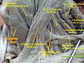
 Sternohyoid muscle - lateral view
 -
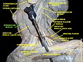
 Sternohyoid muscle - right view
 -
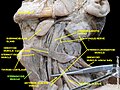
 Sternohyoid muscle
 -
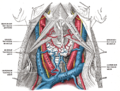
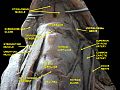 Muscles, nerves and arteries of neck.Deep dissection. Anterior view.


References
![]() This article incorporates text in the public domain from page 393 of the 20th edition of Gray's Anatomy (1918)
This article incorporates text in the public domain from page 393 of the 20th edition of Gray's Anatomy (1918)
- ^ a b c d e Kim, Jong Seung; Hong, Ki Hwan; Hong, Yong Tae; Han, Baek Hwa (2015-03-01). "Sternohyoid muscle syndrome". American Journal of Otolaryngology. 36 (2): 190–194. doi:10.1016/j.amjoto.2014.10.028. ISSN 0196-0709. PMID 25484367.
- ^ a b c d e f g h Standring, Susan (2020). Gray's Anatomy: The Anatomical Basis of Clinical Practice (42th ed.). New York. p. 581. ISBN 978-0-7020-7707-4. OCLC 1201341621.
{{cite book}}: CS1 maint: location missing publisher (link) - ^ Nguyen, John D.; Duong, Hieu (2024), "Anatomy, Head and Neck, Sternohyoid Muscle", StatPearls, Treasure Island (FL): StatPearls Publishing, PMID 31613473, retrieved 2024-02-21
- ^ Chokroverty, Sudhansu (2009-01-01), Chokroverty, Sudhansu (ed.), "Chapter 7 - Physiologic Changes in Sleep", Sleep Disorders Medicine (Third Edition), Philadelphia: W.B. Saunders, pp. 80–104, doi:10.1016/b978-0-7506-7584-0.00007-0, ISBN 978-0-7506-7584-0, retrieved 2020-11-25
- ^ a b Kim, Jong Seung; Hong, Ki Hwan; Hong, Yong Tae; Han, Baek Hwa (2015-03-01). "Sternohyoid muscle syndrome". American Journal of Otolaryngology. 36 (2): 190–194. doi:10.1016/j.amjoto.2014.10.028. ISSN 0196-0709. PMID 25484367.
- ^ Hirano, M.; Koike, Y.; Leden, H. von (1967-01-01). "The Sternohyoid Muscle During Phonation: Electromyographic Studies". Acta Oto-Laryngologica. 64 (1–6): 500–507. doi:10.3109/00016486709139135. ISSN 0001-6489. PMID 6083377.
External links
- Anatomy photo:25:10-0103 at the SUNY Downstate Medical Center - "Nerves and Vessels of the Carotid triangle"
- PTCentral
- v
- t
- e
- Thyrohyoid
- Sternohyoid
- Sternothyroid
- Omohyoid
Portal:
 Anatomy

| Authority control databases |
|
|---|


